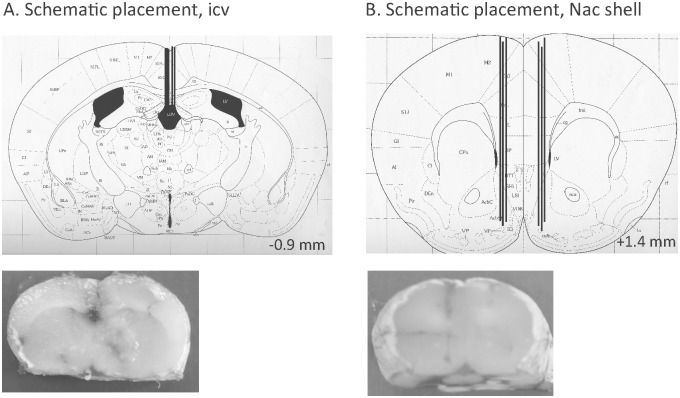
Fig 1. Schematic illustrations of placements.
(A) A coronal mouse brain section showing five representative guide cannula placements (illustrated by vertical lines) aiming at the third ventricle (icv) [25]. In addition, a slice from mouse brain show one representative placement of a guide cannula in the third ventricle (B) A coronal mouse brain section showing six representative probe or guide cannula placements (illustrated by vertical lines) in the nucleus accumbens (NAc) shell [25]. Moreover, a slice from a mouse brain shows one representative placement in the NAc shell. For each brain section only a few representative placements are illustrated, but all other placements targeted the third ventricle or were within the NAc shell. Placements outside these areas were not included in the statistical analysis. The number given in the brain section indicates millimeters anterior (+) or posterior (-) from bregma.

