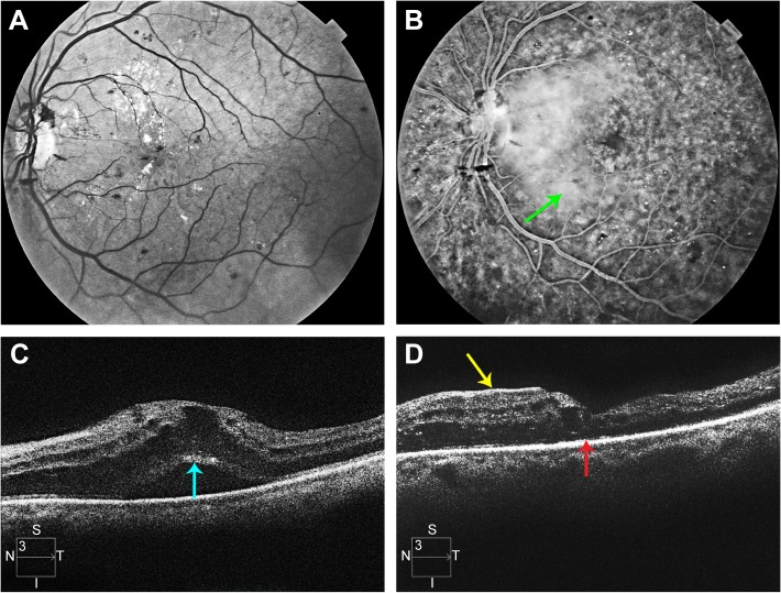
Figure 5.
Multimodal imaging of patient described in case 2.
Notes: (A) Monochromatic fundus photograph of the left eye of the patient described in case 2. (B) Late phase frame of the fluorescein angiogram. The green arrow indicates profuse leakage of fluorescein throughout the macula. (C) Preoperative SD-OCT scan of the left eye. The turquoise arrow indicates the photoreceptor layer with an absent EZ line. (D) 12-month postvitrectomy SD-OCT. The red arrow indicates an absent EZ line. The yellow arrow indicates a nasal ERM that does not cover the fovea.
Abbreviations: SD-OCT, spectral domain optical coherence tomography; EZ, ellipsoid zone; ERM, epiretinal membrane.

