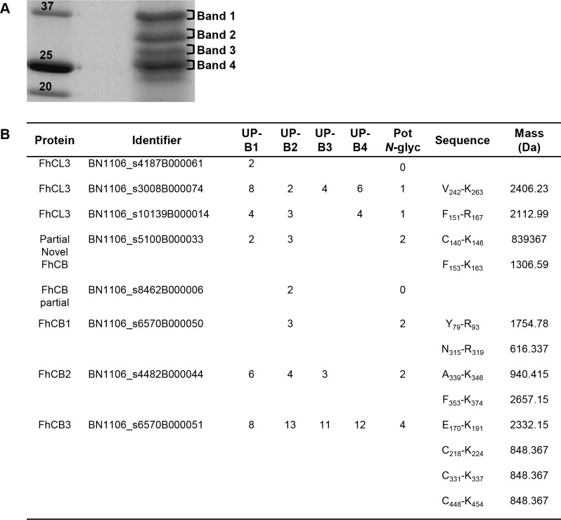
Fig 5. Identification of F. hepatica cathepsins in NEJTeg.
NEJTeg was loaded in SDS-PAGE and stained with Coomassie Blue in order to stain protein bands (A). Bands 1 (B1), 2 (B2), 3 (B3) and 4 (B4), were excised, tryptic digested and identified with LC-MS/MS analysis. Potential N-glycosylation sites (Pot N-gly) and monoisotopic mass of the tryptic peptides containing glycosylation were predicted (B). UP (unique peptide).

