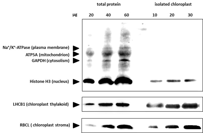
Fig 4. Immunological estimation of contamination in the isolated chloroplasts.
Immunoblot analyses were performed on dilution series of total protein extract (left) and isolated chloroplasts (right). The indicated amounts of each protein were loaded, and the blots were probed with antibodies against proteins from various different subcellular compartments, as indicated. ATP5A, ATP synthase subunit α; GAPDH, glyceraldehyde-3-phosphate dehydrogenase; LHCB I, Light-harvesting chlorophyll protein complex II subunit B1; RBCL, large subunit of Rubisco.

