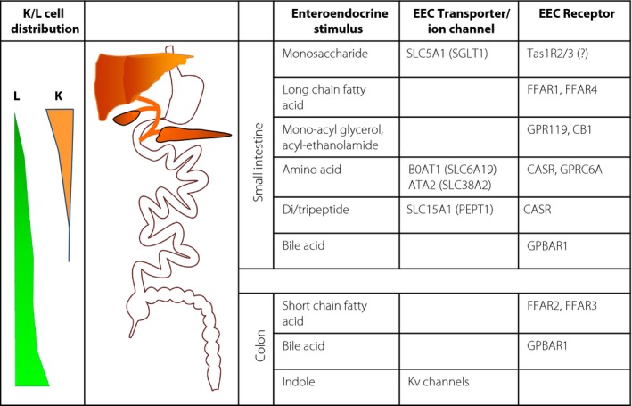
Figure 1.

L‐ and K cell distribution and stimulus detection machinery. The majority of K cells are more proximally located than L cells. Fasting and postprandial glucose‐dependent insulinotropic polypeptide (K cells) and glucagon‐like peptide‐1 (L cells) secretion likely reflect the dynamic gradient of different intestinal stimuli along the gut. Physiological activation of the L‐ and K cell detection pathways is shown, involving transporters/ion channels (linked to altered cellular electrical activity) and G‐protein‐coupled receptors, differing between the small intestine and colon. ATA2, amino acid transporter A2 (solute carrier [Slc] Slc38A2); BOAT1, system B(0) neutral amino acid transporter AT1 (Slc6A19); CASR, calcium‐sensing receptor; CB1, cannabinoid receptor 1; EEC, enteroendocrine cell; FFAR, free fatty‐acid receptor; GPBAR1, G‐protein coupled bile‐acid receptor 1; GPR119, G‐protein coupled receptor 119; GPRC6A, G‐protein coupled receptor classC 6A; Kv‐channels, voltage gated potassium channels; SGLT1, sodium‐coupled glucose transporter 1 (Slc5A1); Tas1R, taste receptor type 1.
