
Figure 2.
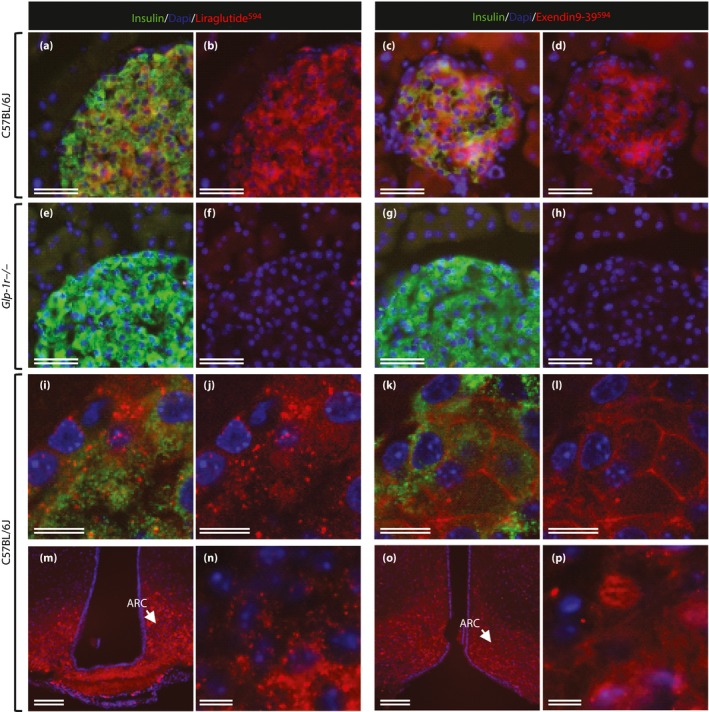
Distribution of liraglutide594 or exendin(9‐39)594 in the pancreas and brain. (a–l) Representative images of mouse islets stained with Hoechst nuclear stain (blue), insulin (green) and liraglutide594/exendin(9‐39)594 (red). (a, c, e, d) Insulin‐ and liraglutide594/exendin(9‐39)594‐positive cells. (b, d, f, h) The same images as in (a, c, e, d) with only Hoechst and liraglutide594/exendin(9‐39)594 signal. (a–d) In C57BL/6J mice, both liraglutide594 and exendin(9‐39)594 were detected in cells expressing insulin; (e–h) however, in mice lacking a functional glucagon‐like peptide‐1 receptor, no liraglutide594 or exendin(9‐39)594 signal could be detected in insulin expressing β‐cells. (i, j, n) High‐magnification images showed that liraglutide594 was internalized and the fluorescent signal was located in the cytoplasm, (k, l, p) while exendin(9‐39)594 remained at the plasma membrane. In the brain, (m, n) liraglutide594 had access to arcuate nucleus (ARC), in which it bound the glucagon‐like peptide‐1 receptor and internalized, (o, p) whereas exendin(9‐39)594 labeled the same population of cells, but without internalization. Scale bars, 100 μm (m, o), 50 μm (a–h), 10 μm (i–l, n, p). ©American Society for Clinical Investigation and reproduced from Secher et al.12 with permission.