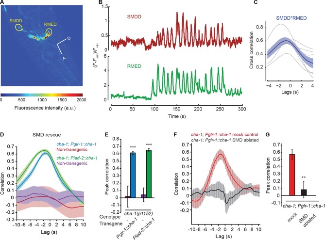
Figure 3. The head bending-correlated calcium activity of RME is regulated by the cholinergic signal from SMD.
(A) Volumetric view of a 3-D image stack from an animal expressing Pglr-1::GCaMP3 and Punc-25::GCaMP3. A, anterior; D, dorsal; circles highlight cell bodies; a.u., arbitrary unit. (B) Sample traces of calcium dynamics in SMDD and RMED during head movement. (C) Cross-correlation between calcium activities in SMDD and RMED. n = 6 animals, faint lines indicate the results from individual animals, the thick line indicates the mean value and the shade indicates SEM. (D, E) Expressing a wild-type cha-1 cDNA in SMD neurons (Pglr-1::cha-1 or Plad-2::cha-1) rescues the head-bending correlated calcium activity in RME. Peak correlation is the highest correlation within the 1 s time window centered on the peak correlation of the wild-type control in Figure 2A and B. (F, G) Ablating SMD in the transgenic animals that express the wild-type cha-1 cDNA with the glr-1 promoter in the cha-1(p1152) mutant animals abolishes the rescuing effect on the head-bending correlated calcium activity in RME. Peak correlation is the highest correlation within the 1 s time window centered on the peak correlation of the mock control. For (E and G), transgenic animals are compared with non-transgenic siblings with student’s t-test. For (D-G) ***p<0.001, **p<0.01, n ≥ 5 animals each, Mean ± SEM; while similar effects were usually observed in more than one transgenic lines, the effect of each transgene is reported with the results from one transgenic line.

