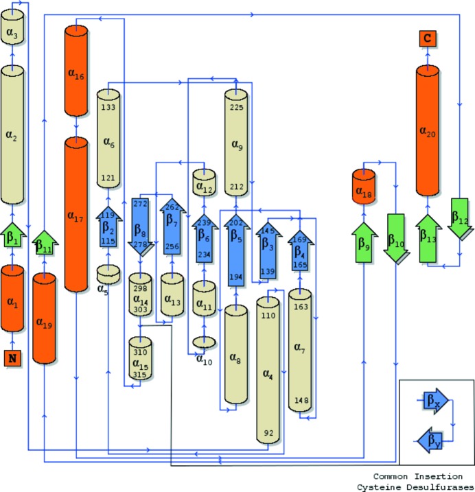
Figure 1.
Topology diagram of secondary-structure elements in PvdN. Small domain components are shown with orange α-helices and green β-sheets, while large domain elements have wheat α-helices and blue β-sheets. Residue numbers are labeled for the secondary-structure elements detailed in the text. The inset displays a common insertion in the cysteine desulfurase family. The diagram was created using the PDBSum server at https://www.ebi.ac.uk/thornton-srv/databases/pdbsum/Generate.html (Laskowski, 2009 ▸).

