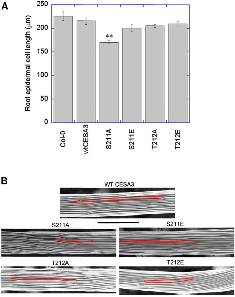
Figure 2.
Root epidermal cell lengths (A) and scanning electron microscope images of etiolated hypocotyls (B). A, Seedlings grown for 7 d on 0.5× MS plates were stained with 1 μg/mL of propidium iodide in water for 4 min. Images of epidermal cells, located in the approximate middle of roots, were captured by a spinning-disk confocal microscope. **P < 0.001. Student’s t test (means ± ses, n = 60-90 cells from 4 to 6 different roots). B, Images of 5-d-old hypocotyls were obtained by using an environmental scanning electron microscope. Bar = 500 μm.

