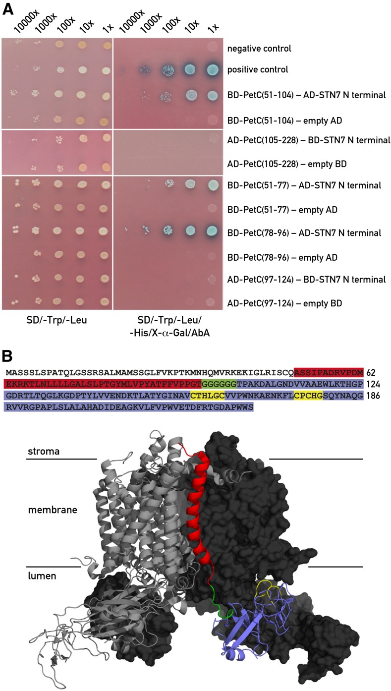
Figure 5.
Interaction between STN7 and Rieske protein. A, Yeast two-hybrid screens with the N-terminal domain of STN7 (residues 44–91) and subdomains of PetC (Rieske protein of the Cytb6f complex). The STN7 and Rieske fragments were either used as bait (BD) fused to the GAL4-DNA binding domain or as prey (AD) fused to the GAL4 activation domain in the yeast two-hybrid assays. This switch between prey and bait had to be performed because of autoactivation of the reporter gene by some of the constructs. Left panel shows the growth test on permissive medium lacking Trp and Leu. Right panel shows the same clones on selective medium lacking Trp, Leu, and His in the presence of aureobasidin A (AbA). The PetC regions are indicated by the residue numbers. Negative and positive controls are described in “Materials and Methods”. B, Summary of the yeast two-hybrid results. The domain of PetC interacting with the N-terminal end of STN7 is highlighted in red. PetC domains giving negative results in the two-hybrid system are highlighted in blue, the hinge region of PetC is in green, and the Cys ligands of the 2Fe-2S center are in yellow. The transit peptide of Pet C is not colored. The structure of the PetC dimer within the Cytb6f complex from C. reinhardtii (Stroebel et al., 2003) is shown in the lower part. The interacting domain is shown in red in the right monomer and the different regions of PetC are shown with the same colors as in the PetC sequence above.

