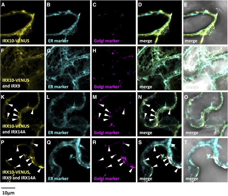
Figure 7.
Fluorescent localization of asparagus IRX10-VENUS proteins expressed in N. benthamiana leaves with or without IRX9/IRX14A. Fluorescence images show IRX10-VENUS (yellow) expressed in N. benthamiana leaves alone (A), with IRX9 (F), with IRX14A (K), or with both IRX9 and IRX14A (P). B, G, L, and Q show the ER marker SP-AtWAK2-CFP-HDEL (cyan). C, H, M, and R show the Golgi marker GmMan1(1-49)-RFP (magenta). D, I, N, and S show the merged images, and E, J, O, and T show the merged images overlaid with the transmitted light images. Golgi localization of IRX10-VENUS is marked with white arrowheads. Overlapping signal is artificially colored white.

