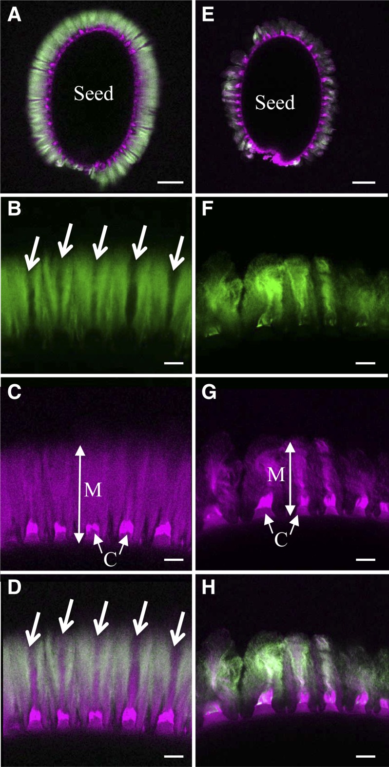
Figure 6.
Labeling of xylan in adherent mucilage released from wild-type and mum5-1 seeds. Confocal microscopy optical sections show adherent mucilage released from mature imbibed seeds after labeling of xylan epitopes with AX1 antibody (green) or staining of cellulose with Pontamine Fast Scarlet 4B (magenta). A to D, Wild-type Col-2 seeds. E-H, mum5-1 seeds. A and E show whole seeds, and B, C, and D or F, G, and H show higher magnifications of mucilage from the same seeds, respectively. A, D, E, and H show composite images of double labeling with Pontamine and AX1 antibody. Arrows indicate zones in adherent mucilage that are not labeled with AX1. C, Columella; M, adherent mucilage. Bars = 100 µm (A and E) and 20 µm (B–D and F–H).

