Figure 1.
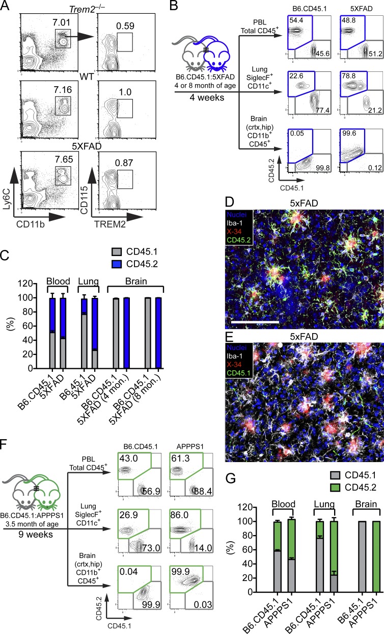
Lack of monocyte contribution to amyloid-associated microglia. (A) Surface expression of TREM2 among Ly6C+CD11b+CD115+ blood monocytes in WT and 5XFAD mice. Trem2−/− mice were used as negative controls. (B–E) Parabiosis was performed by joining blood circulation of 4- or 8-mo-old 5XFAD with age matched B6.CD45.1 congenic mice for 4 wk. (B) CD45+ blood leukocytes, CD11c+SiglecF+ lung alveolar macrophages, and brain CD11b+F4/80+ microglia from parabionts were analyzed by flow cytometry. (C) Frequencies of CD45.1+ and CD45.2+ cells were compared in parabiotic partners. (D and E) Representative images of brain sections of an 8-mo-old 5XFAD parabiont stained with X-34 (red) for Aβ plaques and Iba-1 (white) for microglia. Contribution of host- and donor-derived microglia was examined using antibodies specific for CD45.2 (green; D) and CD45.1 (green; E). Nuclei were visualized with To-pro3 (blue). Bar, 100 µm (F and G) Parabiosis was performed using APPPS1-21 mice and age-matched B6.CD45.1 congenic mice for 9 wk. Parabionts were analyzed as described above. Data represent a total of five to seven mice per group in A–E and four mice per group in F and G.

