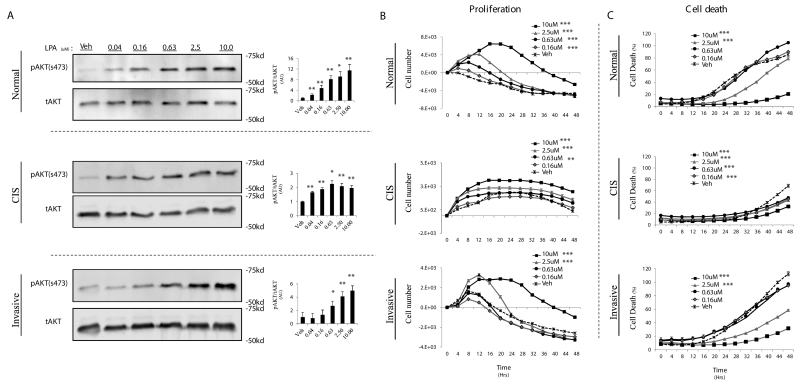
Figure 2.
LPA initiates a proliferative and survival signal for TAg MECs. A, TAg cell lines were serum starved for 8hr, followed by a 10 minute treatment with the indicated concentration of LPA. AKT phosphorylation at serine 473 (pAKT) levels was quantified by densitometry analysis of Western blots and normalized to total AKT levels (right, pAKT/AKT) (A). TAg cell lines were cultured without serum +/− the indicated concentration of LPA and then cell proliferation (B) and cell death (C) were measured using the Incucyte™ live content imaging system. LPA was only added at the initiation of the experiment. Cell numbers were calculated from confluence values (described in methods). Change in cell number was calculated by subtracting the initial cell count at treatment time=0 from all subsequent cell counts. Error bars indicate standard error of the mean. Statistics were determined from 3 independent experiments. In all panels; *, p≤ 0.05; **, p≤0.01; *** p≤0.001.

