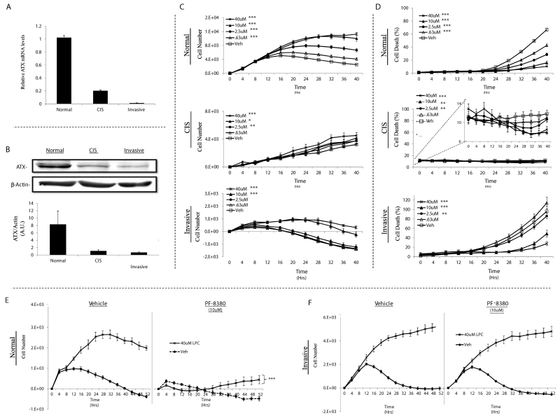
Figure 5.
LPC supports the proliferation and survival of TAg MECs and requires LPAR signaling in normal but not invasive TAg MECs. Relative expression of ATX (Enpp2) mRNA transcripts (A) and protein (B) in TAg cell lines. TAg cell lines were cultured without serum +/− the indicated concentration of LPC and then cell proliferation (C) and cell death (D) were monitored using the Incucyte™ live content imaging system. Normal (E) and invasive (F) TAg cell lines that were cultured without serum and in the presence or absence of 40uM LPC were treated with 10uM PF-8380 (ATX antagonist) or vehicle. The cells were then placed in the Incucyte™ live content imaging system to measure proliferation for 52 hours. Statistics were determined from 3 independent experiments. Error bars indicate standard error of the mean. Panels C - E; *, p≤0.05; **, p≤0.01; *** p≤0.001 compared to vehicle (control).

