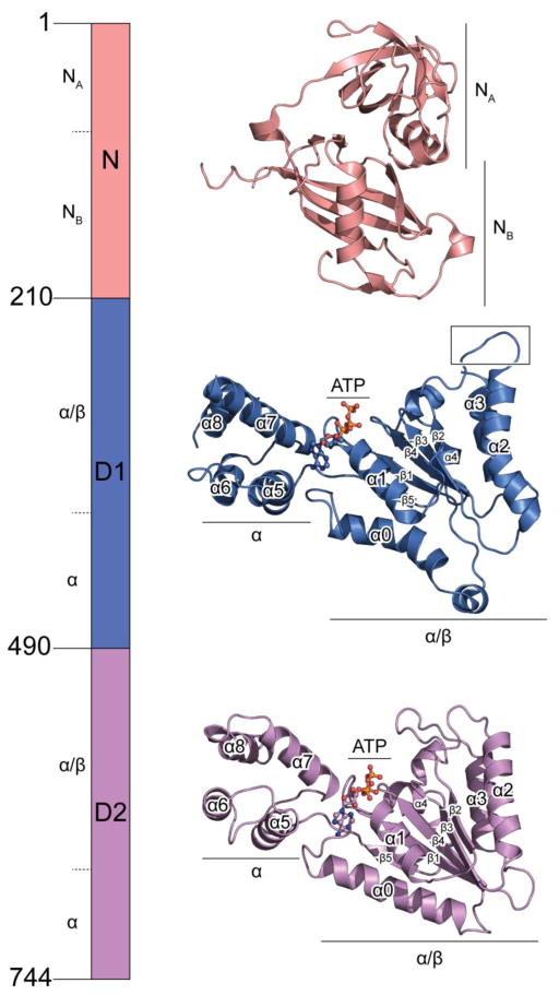
Fig. 1. Domain architecture of NSF.
Crystal structures of the N domain (PDB accession code: 1qcs), and the D2 domain (PDB accession code: 1nsf) are shown. The structure of the D1 domain is from the cryo-EM structure of NSF in the ATP state (PDB accession code: 3j94). The structures are shown in scale, with subdomains marked. D1 and D2 domains are aligned in a similar orientation with all the conserved secondary structure elements of the AAA+ domain (defined in [22]) labeled. Compared to the D2 domain, the D1 domain has a characteristic bent α2 helix.

