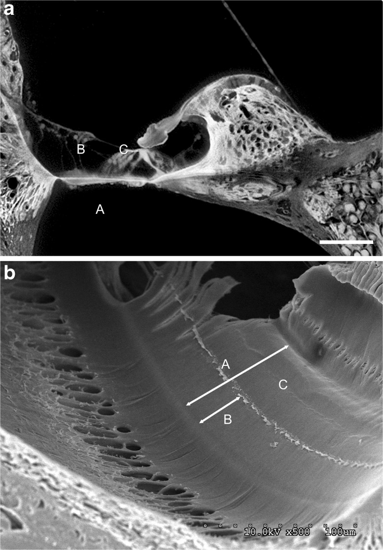
FIG. 1.
a An sTSLIM cross section of the scala media from a cellularized mouse cochlea showing the basal turn. “A” marks the width of the basilar membrane (83 μm). “B” marks the distance (36 μm) from the lateral edge of the basilar membrane to the beginning of the Boettcher cells. “C” marks the distance (47 μm) from the medial edge of the basilar membrane to the beginning of Boettcher cells. Bar = 50 μm. b An SEM view of the scala media in a decellularized cochlea showing the basal turn. “A” marks the width 78 (μm) of the basilar membrane. “B” marks the distance (32 μm) from the lateral edge of the basilar membrane to the structure. “C” marks the distance (43 μm) from the structure to the medial edge of the basilar membrane. Bar = 100 μm.

