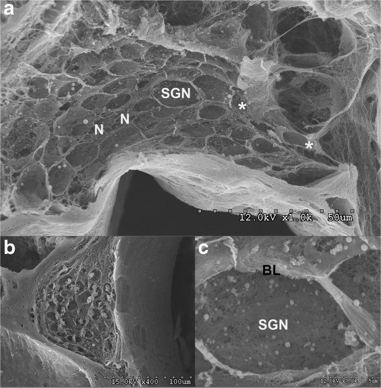
FIG. 10.
a–c SEM views of Rosenthal’s canal and the spiral ganglion. a Numerous empty openings that previously housed the spiral ganglion neurons (SGN) are evident (15 μm in diameter) as well as openings for nerve fibers (N) (3 μm in diameter) and where the cell bodies extended into the nerve processes (*). b In a deoxycholate-treated cochleas, the contents of the spiral ganglion neurons were not completely extracted by the detergent therefore a residue remains. c Higher magnification view of an opening that previously contained a spiral ganglion neuron (SGN) surrounded by basal lamina material (BL) and additional fibrillar ECM material.

