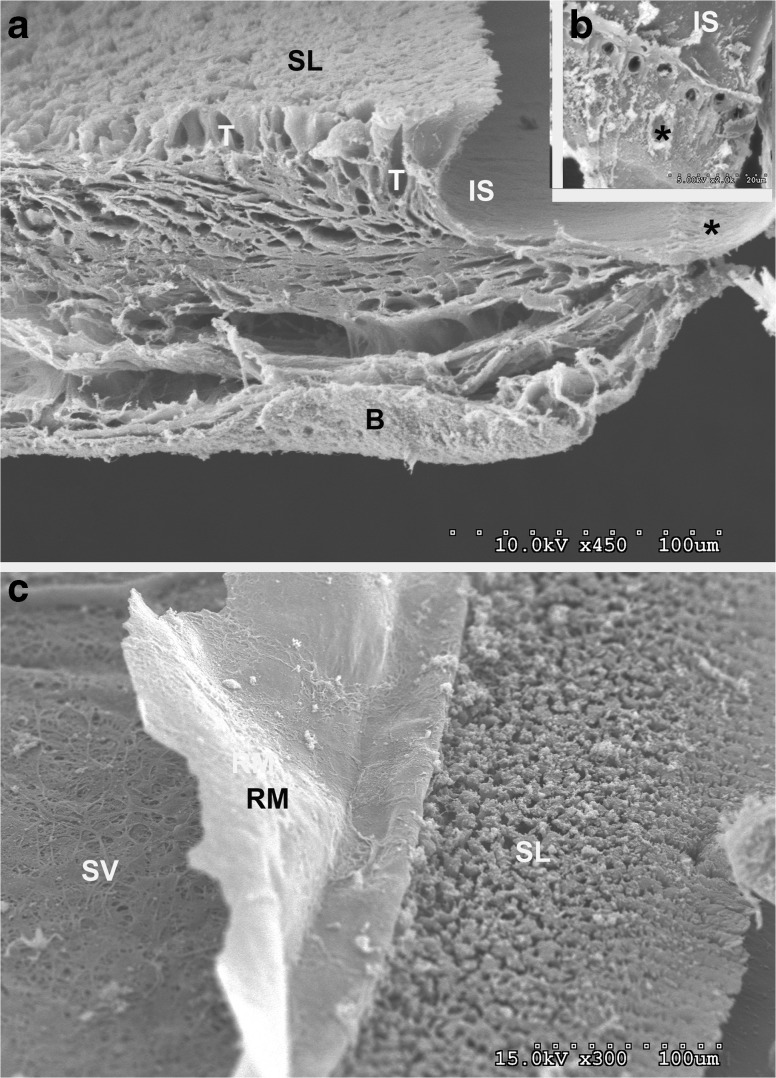
Fig. 12.
a Higher magnification SEM view of the basal end of the scala media showing the bone (B), spiral limbus (SL) with spaces where the interdental T cells occupied (T), inner sulcus (IS), and inner hair cell region near the habenula perforata (*). b View of inner sulcus cell region (IS), the openings of the habenula perforate and septa-like features (*) at the basal border of the cells at the inner hair cell region. c Lower magnification view of the bone of the scala vestibule (SV) showing fibers and openings, the basal lamina of Reissners membrane (RM), and the apical portion of the spiral limbus (SL) showing openings between the heads of the spiral limbus plates.

