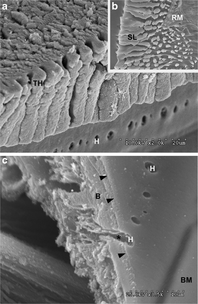
FIG. 3.
a SEM view of the spiral limbus in a decellularized cochlea showing basal lamina coated columns of ECM, the teeth of Huschke (TH) and the habenula perforata (H) with a diameter of approximately 2 μm. b The spiral limbus (SL) at the basal end of the cochlea where there is extensive basal lamina covering heads of the smaller pillars. A small portion of the basal lamina of Reissner’s membrane (RM) is also present. c A fracture through the osseous spiral lamina and a habenula perforata (H) showing the extension of the channels where the nerves travel through the habenula (*). The basilar membrane is shown on the lower right (BM), and the arrowheads indicate the coating of basal lamina material over the bone (B).

