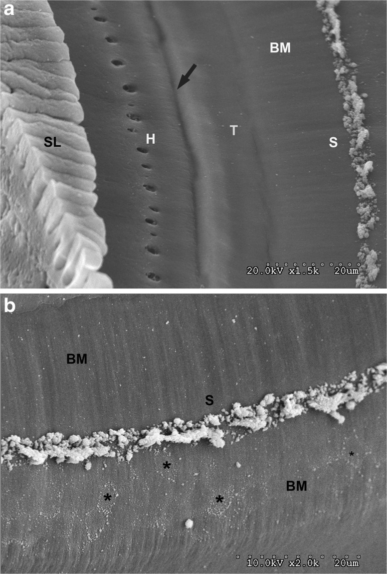
FIG. 5.
a SEM of a decelluarized cochlea showing the spiral limbus (SL), habenula perforata (H), and the medial beginning of the basilar membrane as indicated by a spirally directed dark line (arrow). Light and darker areas are more lateral (T) where presumably the tunnel cells use to lie, and then, the smooth surface basilar membrane extends to the structure (S) shown in an OTO processed cochlea. b A higher magnification view of the basilar membrane (BM), structure (S), and numerous pentagonal/hexagonal imprints of the Boettcher cells (*) on the side of the structure facing the lateral wall.

