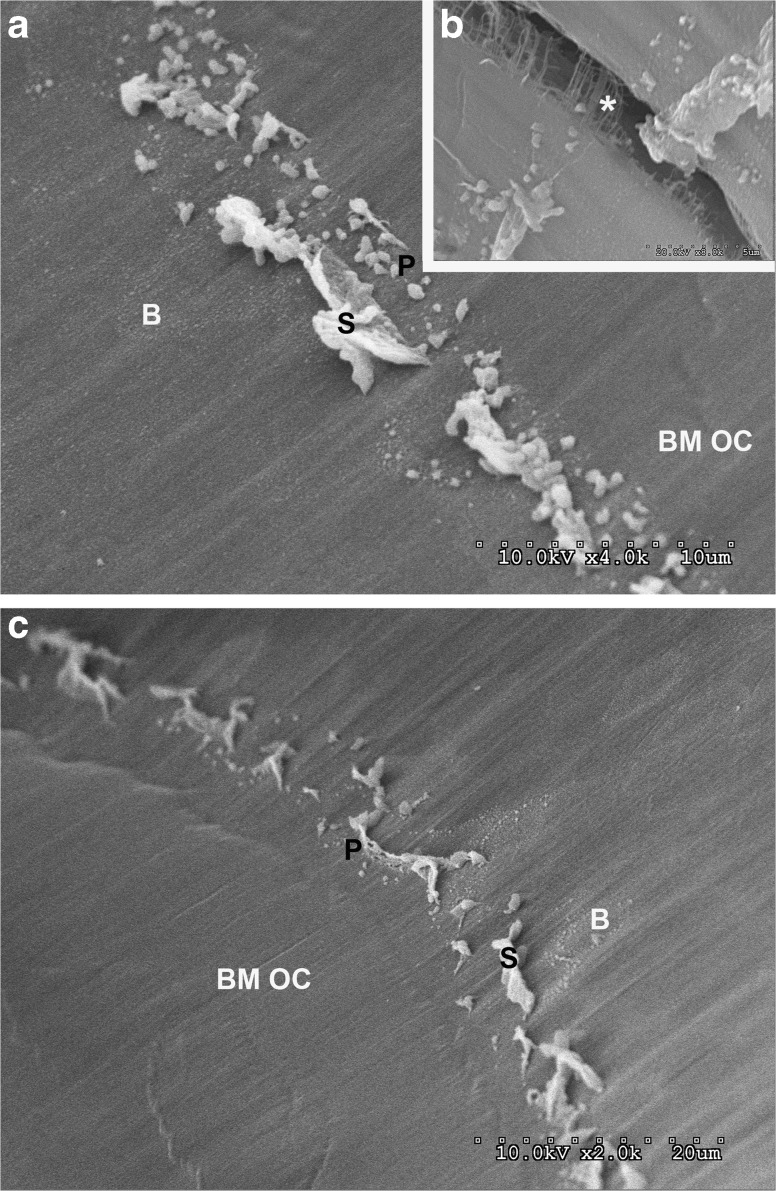
FIG. 6.
a SEM views of the structure from a decellularized cochlea not processed with OTO. It forms a spirally directed, single line of periodic structures. They show septa-like (S) protrusions from the basilar membrane on the Boettcher side (B) of the basilar membrane and particles (P) on their side facing the organ of Corti (BM OC). b Higher magnification insert shows a crack in the basilar membrane and fine filaments extending radially in the deeper layers of the basilar membrane. c Another cochlea showing similar morphology of the structure with a periodic distribution, septa, particles, and imprints. Radial and some spiral striations appear in the basilar membrane.

