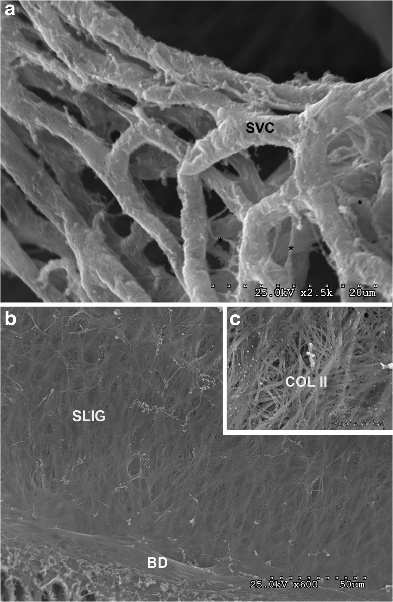
FIG. 8.
a A network consisting of the basal lamina of the capillaries of the stria vascularis (SVC). The capillaries are approximately 3 μm in diameter, branched and are associated with a fine network of thin (0.3 μm) filaments (*). The capillary basal lamina appears to be “free floating” above the spiral ligament and are connected to the lateral wall by the collecting venues and radiating arterioles. b The spiral ligament (SLIG) is composed of a meshwork of filaments that are 5 μm in diameter or smaller. The surface facing the previous stria vascularis is relatively smooth and consists of a loose network of cross-connecting filaments. A band (BD) lies between the spiral prominence and the spiral ligament. c Insert shows details of these filaments which are presumably collagen II (COL II).

