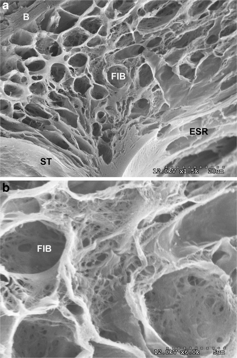
FIG. 9.
This figure shows surface of the scala tympani (ST) on the left, a cross-section of the spiral ligament at the top with openings, which previously contained fibrocytes (FIB) and a portion of the scala media with the external sulcus cell openings. b Shows the filamentous network of the spiral ligament and the openings (FIB) previously occupied by fibrocytes that are surrounded by type II collagen filaments.

