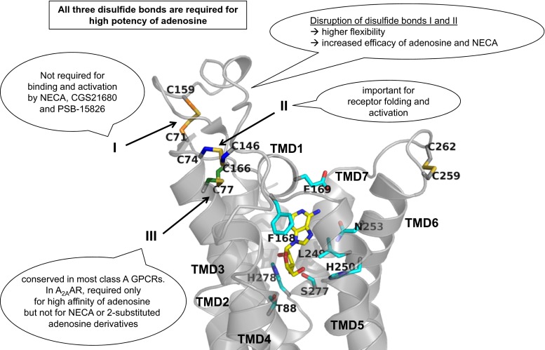
Fig. 6.
Binding modes of adenosine in the A2AAR. Crystallographic binding poses of the endogenous agonist adenosine (represented in stick, carbon atoms in yellow) in the binding pocket of the A2AAR (represented as gray ribbon) are shown. The side chains of important residues in the binding pocket are shown as sticks with carbon atoms in cyan. The cysteine residues involved in disulfide bonds are shown as sticks, and the carbon atoms are color-coded (Cys712.69-Cys15945.43 orange, Cys743.22-Cys14645.30 blue, and Cys773.25-Cys16645.50 green). The disulfide bond number is indicated. The main results are summarized in figure

