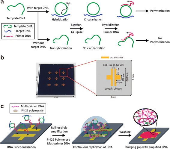
Fig. 1.

Schematic illustration of pathogen DNA detection on Au electrodes. a Synthesis of the closed circular DNA in the presence of target DNA (top). In the absence of target DNA, no circularization occurred (bottom). Template DNA strands in both cases are introduced with thiolated primer DNA to form hybridization. b Digital camera image (left) and detailed illustration (right) of the Au electrodes on Si/SiO2 substrate. c RCA process on Au electrodes with multi-primer DNA (purple). The amplified DNA strands fill the gap between the two electrodes
