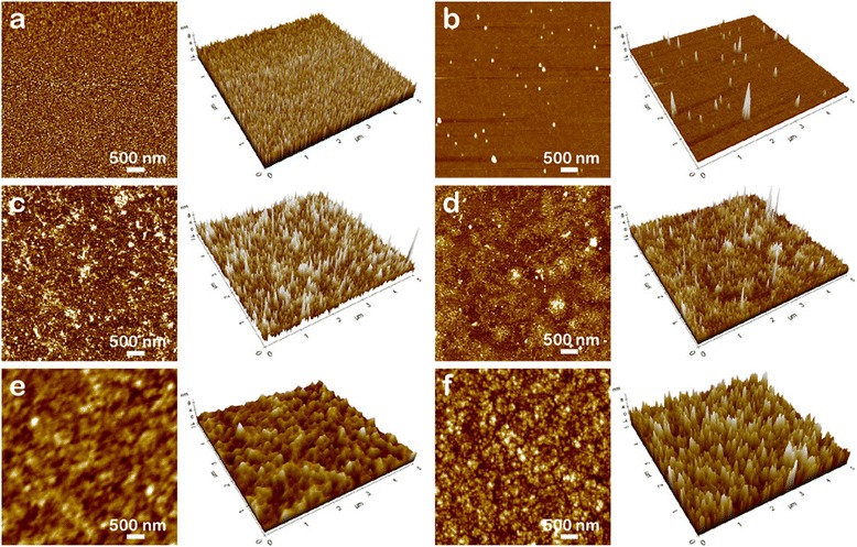
Fig. 2.

AFM images of the surfaces of Au electrodes and the gap between the electrodes. The surface of the Au electrodes (a, c, e) and the gap (b, d, f) between the electrodes (a, b the untreated; c, d treated with 0.05 μM of the target DNA; e, f treated with 1 μM of the target DNA)
