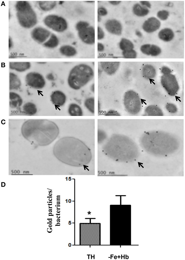
Figure 2.

Immunoelectron microscopy of Spbhp-37 protein. S. pneumoniae cellular cultures were incubated in normal Todd-Hewitt Broth or in medium with Hb as only iron source. Then, localization of Spbhp-37 was analyzed by immunoelectron microscopy. (A) Negative control. Bacteria incubated only with the gold-labeled secondary antibodies. (B) Spbhp-37 in normal medium. (C) Spbhp-37 in medium with Hb as only iron source. Arrowheads indicate the location of Spbhp-37 protein in S. pneumoniae. In right is shown the magnification of cell bacteria to compare expression in both growth conditions. (D) Quantitative analysis of Spbhp-37 expression. Gold particles on bacteria growth under normal conditions (TH) and in medium with Hb as only iron source (-Fe+Hb) were counted n = 25. Data represent mean ± SD of three independent experiments. Asterisk indicates a significant difference (p < 0.05).
