
Figure 3.
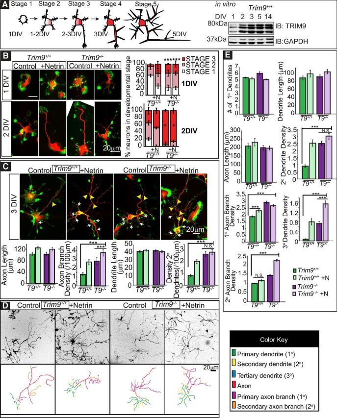
Deletion of Trim9 results in increased dendritic arborization and axon branching in vitro. A, Graphical representation of neuronal stages of morphology. Stage 1 neurons lack neurites. Stage 2 neurons have 1 or more neurites. Stage 3 neurons have a presumed axon at least 2 times the length of other neurites. Axonal and dendritic branches emerge in Stage 4 and Stage 5 neurons, respectively. Western blot of TRIM9 in E15.5 hippocampal neurons in vitro over time. B, Immunofluorescence of Trim9+/+ (T9+/+) and Trim9−/− (T9−/−) dissociated hippocampal neurons at 1 and 2 DIV and quantifications of percentage of total cells per developmental stage, ± SEM from three independent experiments. C, Immunofluorescence of Trim9+/+ and Trim9−/− dissociated hippocampal neurons at 3 DIV and quantifications of average length (μm), average number of neurites, and average branches/100 μm primary neurite length at 3 DIV. Red represents βIII-tubulin. Green represents F-actin (phalloidin). White arrows indicate secondary dendrite branches. Yellow arrows indicate axon branches. D, Immunofluorescence and color-coded tracings of Trim9+/+and Trim9−/− dissociated hippocampal neurons at 5 DIV. Images have been inverted from a grayscale merged F-actin and βIII-tubulin image. E, Quantifications of average length (μm), average number of neurites, and average branches/100 μm primary neurite length for 5 DIV. Error bars indicate SEM. *p < 0.05. **p < 0.005. ***p < 0.0005. NS, Not significant.