Figure 4.
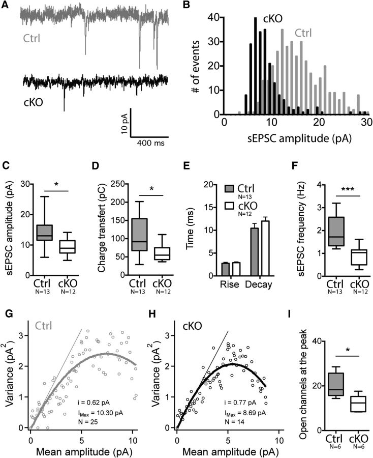
Spontaneous EPSCs are decreased in NMDA-R-deficient iMSNs. A, Representative current traces of sEPSCs recorded from control and cKO mice. B, Distribution of sEPSC amplitudes in a cKO and a control cell showing a significant shift to the left for the cKO neuron. C, D, Summary graph showing a decrease of sEPSC amplitude in cKO mice (C), leading to a reduced charge transfer (D). E, There was no difference in kinetic parameters for either the rise slope or the decay slope. F, sEPSC frequency was decreased in cKO animals. G, H, Peak-scaled nonstationary fluctuation analysis was performed on sEPSC recordings and analysis of representative control and cKO cells is presented. The slope represents the unitary channel current. I, Estimated number of AMPA-R open at the peak is lower in cKO cells.

