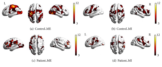
Figure 1.
Brain activation in the control and patient groups under different conditions. (a) Control subjects during motor execution; (b) controls during motor imagery; (c) patients during motor execution; (d) patients during motor imagery. All voxels were significant at p < 0.01, corrected for FDR at the whole-brain level.

