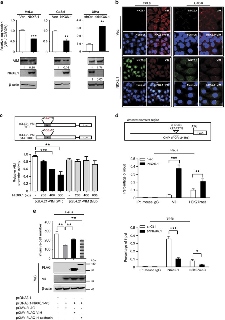
Figure 6.
NKX6.1 represses cancer invasion by decreasing the expression of mesenchymal markers. (a) The expression of vimentin in NKX6.1-expressing cells and NKX6.1-shRNA-knockdown cells was assessed by qRT–PCR (bar graphs) and western blotting (below). The numbers in the western blots represent the ratios of targets to the internal control. (b) IF assays were used to detect vimentin expression in NKX6.1-expressing HeLa and CaSki cells. (c) The upper scheme depicts WT (pGL4.21-VIM) and HDBS-mutated (Mut; pGL4.21-VIM) promoters of the vimentin gene. The activity of different vimentin promoter constructs in HeLa cells was analyzed by a luciferase reporter assay. (d) Chromatin from HeLa cells expressing NKX6.1 or SiHa cells expressing NKX6.1 shRNAs was immunoprecipitated with indicated antibodies and then analyzed by quantitative PCR using vimentin-specific primers. The upper scheme depicts the position of the HDBS within the promoter region of vimentin. (e) HeLa cells were transfected with the indicated combination of vectors, and Matrigel invasion assays were used to analyze the effects on cancer invasion. The data are presented as the mean±s.e. from three independent experiments in triplicates (analyzed by unpaired two-tailed t-test). *P<0.05, **P<0.01 and ***P<0.001.

