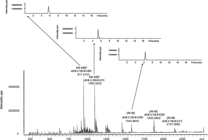
Figure 2. The mass spectrum and its corresponding extracted ion chromatogram (XIC) obtained by UHPLC-ESI-FTICR MS.
The structure of a ganglioside includes a ceramide tail (fatty acid N-linked to a sphingosine) of varying length, saturation and hydroxylation linked to a polar carbohydrate head that contains sialic acid. The gangliosides are grouped according to the number of their sialic acid residues: one (M), two (D), three (T) or four (Q).GM, GD and GT are main gangliosides in brain.

