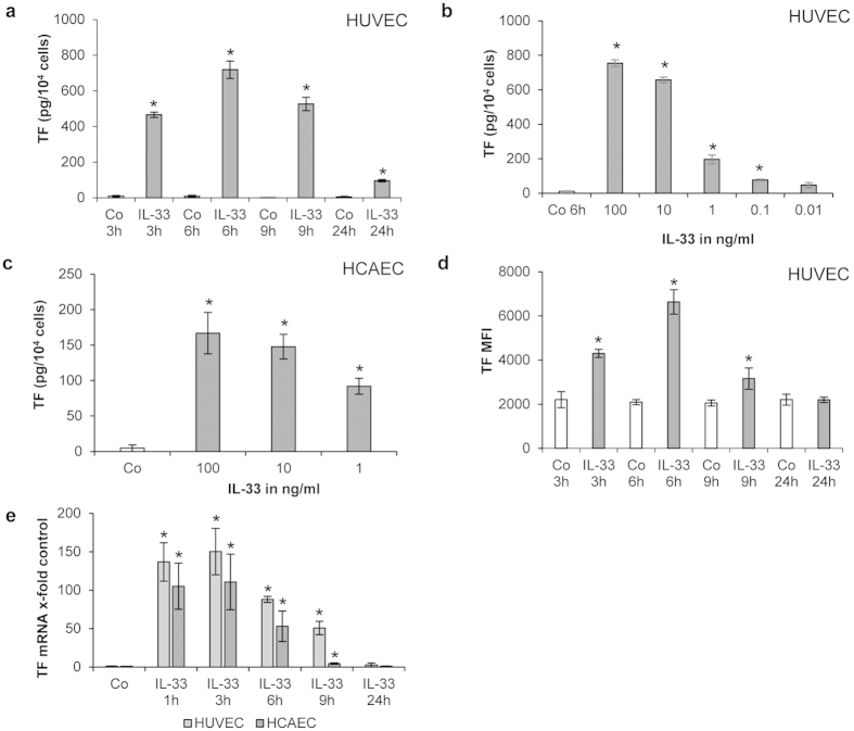
Figure 1. IL-33 increases TF protein and mRNA in human ECs in a time– and concentration-dependent manner.
(a,b) HUVECs were incubated for 3, 6, 9 and 24 hours (h) in the absence (control, Co) or presence of rh IL-33 (100 ng/mL) (a) or HUVECs were incubated for 6 h without (Co) or with rh IL-33 at 100, 10, 1, 0.1 and 0.01 ng/mL (b). Cells were permeabilized with PBS containing 0.1% Triton X-100 and TF protein was determined; (c) HCAECs were incubated for 3 h without (Co) or with rh IL-33 at 100, 10, and 1 ng/mL. Cells were permeabilized with PBS containing 0.1% Triton X-100 and TF protein was determined. (d) HUVECs were incubated for 3, 6, 9 and 24 h without (Co) or with 100 ng/mL IL-33. TF expression at the cell surface was measured by means of flow cytometry; (e) HUVECs and HCAECs were incubated for 1, 3, 6, 9 or 24 h in the absence (Co) or presence of IL-33 (100 ng/mL). mRNA was prepared and Real Time PCR was performed. Values are given in pg/104 cells (a,b,c), in mean fluorescence intensity (MFI) (d) or as TF/GAPDH mRNA x-fold change from respective control, which was set as 1 (e) and represent mean values ± standard deviation (SD) of three independent determinations. *p ≤ 0.05 compared to respective control.

