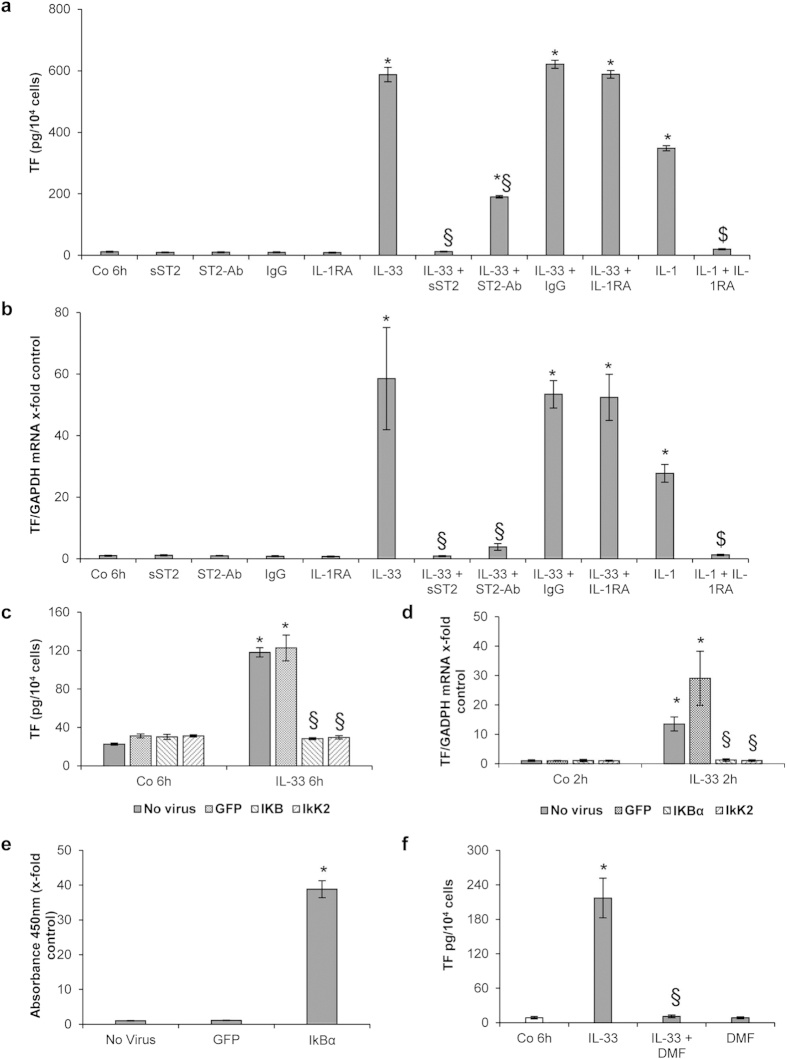
Figure 2. IL-33-induced TF expression is ST2- and NF-κB-mediated in HUVECs.
(a,b) HUVECs were left untreated (Co), or incubated for 6 h with rh IL-33 (10 ng/mL) in the presence or absence of rh ST2/IL-1 R4 Fc chimera (sST2; 5 μg/mL), rh immunoglobulin G1 Fc (IgG; 5 μg/mL) or IL-1 receptor antagonist (IL-1RA; 10 μg/mL). In addition, IL-1β (100 U/mL) was added to the cells for 6 h with or without 10 μg/mL IL-1RA. Additionally, cells were pre-incubated for 30 min with 1 μg/mL human ST2/IL-1R4 antibody (ST2-Ab), before addition of 10 ng/mL IL-33. Cells were permeabilized with PBS containing 0.1% Triton X-100 and TF protein was determined (a), or mRNA was prepared and Real-Time PCR was performed (b). (c–e) HUVECs were left uninfected or were infected with adenoviral vectors for overexpression of IκBα (AdV-IκBα) or a dominant negative form of IκB kinase 2 (AdV-dnIkK2) or with a control adenovirus (AdV-green fluorescent protein (GFP)) for 4–6 h. 48 h post infection cells were treated with IL-33 (1 ng/mL) for 2 and 6 h whereas control cells were left untreated. Cells were permeabilized with PBS containing 0.1% Triton X-100 and TF protein (c) or IkBα protein (e) was determined, or Real-Time PCR for TF and GAPDH mRNA was performed (d).(f) HUVECs were treated with medium alone (control, Co), IL-33 alone (10 ng/mL), dimethyl fumarate (DMF) at 100 μM alone or together with IL-33 for 6 h. Cells were permeabilized with PBS containing 0.1% Triton X-100, and TF protein was determined. Values are given in pg/104 cells (a,c,f) as x-fold absorbance at 450 nm as compared to control (no virus; (e)) or as TF/GAPDH mRNA x-fold change from respective control, which was set at 1 (b,d) and represent mean values ± SD of three independent determinations. *p ≤ 0.05 compared to respective control; §p ≤ 0.05 compared to respective IL-33-treated cells; $p ≤ 0.05 compared to respective IL-1β-treated cells.

