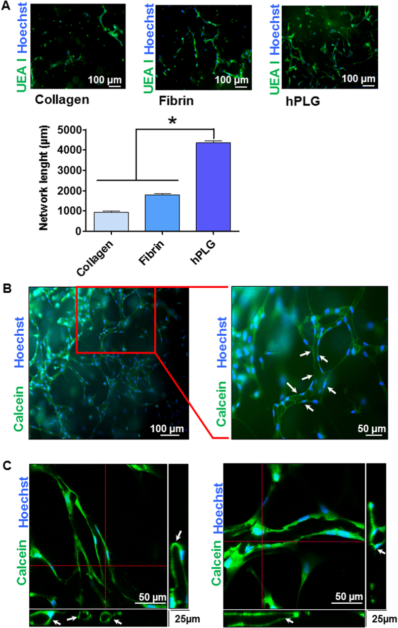
Figure 4. Detection of a lumenized vascular network by hPLG encapsulated ECFCs using epifluorescence and confocal microscopy.
(A) ECFCs were encapsulated in collagen I, fibrin or hPLG and cultured for 3 days at 37 °C. Constructs were fixed and stained with FITC-conjugated Ulex europaeus lectin (FITC-UEA I) which binds to α-L-fucose-containing glycocompounds on the surface of endothelial cells and Hoechst 33342 to localize cell nuclei. Total length of the capillary network in the different conditions was measured using Angiogenesis Analyzer plugin of ImageJ 1.49 v. As normal distribution and equal variance of the samples were confirmed using the Shapiro-Wilk normality test and Bartlett’s homoscedasticity test (Supplementary Table 2), statistical significance was tested using one way ANOVA with Bonferroni post-test (*P ≤ 0.05, n = 6). hPLG-encapsulated ECFCs were live stained after 3 days of the start of the experiment. Fluorescent calcein AM was used to stain the cytoplasm of viable cells, while Hoechst 33342 was utilized to localize cell nuclei. Images show multiple ECFCs organized into primitive vascular structures with a lumen (white arrows). Epifluorescence images were obtained using a Leica DMI4000B microscope (objective: N Plan 10X/0.25) (B), whereas confocal microscopy was performed with a LSM 510 META confocal microscope (objective: EC Plan Neofluar 10X/0.30 M27) (C).

