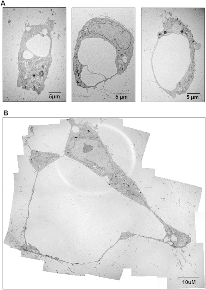
Figure 5. Detection of a lumenized vascular network by hPLG encapsulated ECFCs using transmission electron microscopy (TEM).
TEM was used to assess whether the tubular structures developing in ECFCs cultured in hPLG have a lumen and display capillary morphology. Different stages of lumen formation are shown: from vacuole formation to lumens delimited by single cells (A) to multicellular lumen delimitation (B). Images are representative of three independent experiments.

