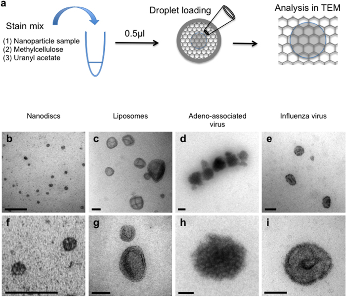
Figure 1. Nanoparticles in methylcellulose films imaged in TEM.
(a) The stain mix contained 1.5 μl of 1% methylcellulose, 7.5 μl of nanoparticle suspension and 1 μl of 0.3% uranyl acetate (uranyl acetate was added after dialysis if required; see text and Fig. 3). 0.5 μl was loaded onto a standard pioloform coated EM grid support and allowed to dry before examination in the TEM. (b–i) A range of nanoparticles imaged at low magnification (b–e) and high magnification (f–i). Scale bars (b–e) 100 nm and (f–i) 50 nm.

