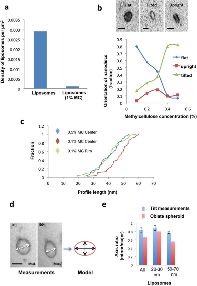
Figure 2. Methylcellulose restricts binding to the substratum, allows freedom of orientation and prevents substantial collapse of nanoparticles.
(a) Binding to substratum. Liposomes were incubated in 1% methylcellulose or water before adsorbing the mix to an EM grid, contrasting, and density of liposomes determined. (b) Orientation. 12 nm diameter nanodiscs were mixed with methylcellulose solution varying between 0.1% or 0.5% before adding uranyl acetate. 0.5 μl droplets were loaded onto EM grids and dried before classifying orientation by TEM (flat, tilted and upright). Nanodiscs become progressively tilted with increasing concentration of methyl cellulose. Bars, 7nm. (c) Gold nanorods were mixed with 0.1% or 0.5% MC. 0.5 μl drops were loaded onto EM grids and nanorods situated in either the rim or center were measured. Data are presented as cumulative fractions. Nanorods in the thinnest films (0.1% MC center) were longer compared to those in thicker films at the 0.1% MC rim (Kolmogorov-Smirnov test, P 0.001; n 45 and 52 data points respectively) or the 0.5% MC center (Kolmogorov-Smirnov test, P 0.01; n 45 and 38 data points respectively). The data are consistent with increased freedom to rotate in thicker MC films. (d,e) Tilt analysis of vesicle compression in methylcellulose films. Vesicles containing (MscL) pore protein were embedded in methyl cellulose as described in the text and the degree of flattening (oblate spheroid; model) estimated from minor and major axes measured after 50° specimen tilt. Data are from the whole population (All), n = 38, 20–30 nm vesicles, n = 16 and 50–70 nm vesicles, n = 10. Bar = 50 nm.

