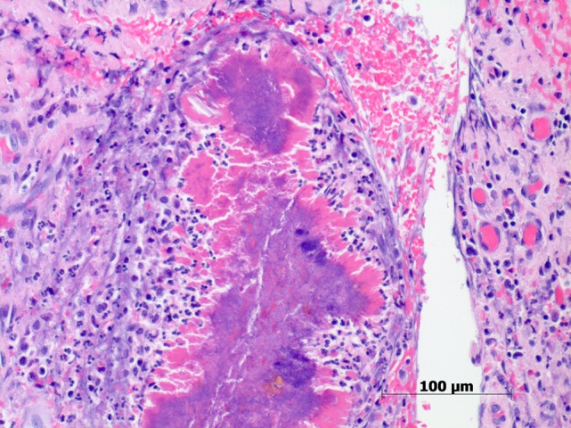
Figure 4:

Histopathology of appendix for Case 2. Sulfur granules showing clusters of Gram-positive filamentous non-spore forming actinomyces with adjacent neutrophilic infiltrate. The filaments are surrounded by eosinphilic proteinaceous material which represents a host reaction (haematoxylin–eosin stain, original magnification ×200).
