
Fig. 4.
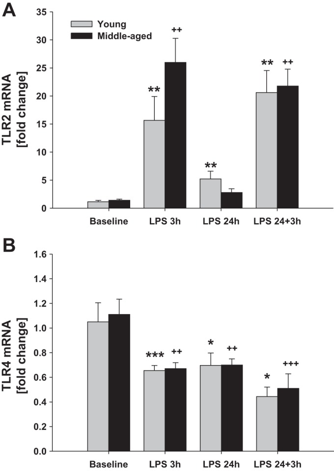
Microglial Tlr2 and Tlr4 mRNA levels after intraperitoneal LPS injection. Young adult and middle-aged mice were injected with either PBS (baseline) or LPS (5 mg/kg ip) at times 0 and 24 h. Microglia were immunomagnetically isolated at 3 and 24 h after the 1st LPS injection and 3 h after the 2nd LPS injection (24+3 h). qRT-PCR was used to evaluate the expression of Tlr2 (A) and Tlr4 (B). 1 symbol, P < 0.05; 2 symbols, P < 0.01; 3 symbols, P < 0.001; *time point vs. baseline (0 h) in young mice; +time point vs. baseline (0 h) in middle-aged mice.