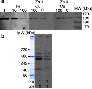
Fig. 5.

Fe and Zn-dependence of OtFea1 expression, and OtFea1 iron band on native PAGE. a O. tauri cells were grown for one week in Mf medium containing either 1, 10 or 100 nM Fe (as ferric citrate) or in Mf medium containing 1 nM Fe and different concentrations of Cu (0 or 100 nM CuSO4) and Zn (0 or 1 μM ZnCl2). Whole-cell extracts were prepared as described in the methods, and proteins (25 μg/lane) were separated by SDS-PAGE before immunoblotting with an anti-OtFea1 primary antibody. b Autoradiograph showing the main iron bands after short-term iron loading of the cells and protein separation by native PAGE. O. tauri cells were grown for five days in Mf medium containing 1 nM Fe and either 0 or 1 μM Zn, and then incubated for 3 h with 5 μM 55Fe(III)-citrate as described in the methods. Whole-cell extracts were obtained and subjected to native PAGE (25 μg/lane). After autoradiography, iron-containing bands were analyzed by mass spectrometry. OtFea1 was found in the band indicated by a star (*)
