
Fig. 7.
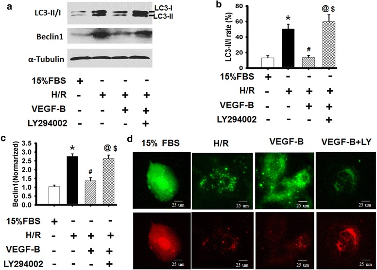
VEGF-B inhibited H/R-induced autophagy through PI3/Akt signaling. a LC3-II/I expression was detected using western blot, as described in the “Methods” section (a), and quantified via normalization to a-tubulin (b, c). *P < 0.05 vs. 15 % FBS-treated cells; # P < 0.05 vs. H/R-treated cells; @ P < 0.05 vs. 20 ng/ml VEGF-B-treated cells; $ P > 0.05 vs. H/R-treated cells, n = 6. d Representative images showing LC3 staining in different groups of H9c2 cells infected with Ad-GFP-RFP-LC3 for 48 h, n = 6