
Fig. 3.
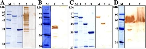
SDS-PAGE and immunoblot analysis of recombinant and native BcMSA1 and BcSA1. a The 10 % SDS-PAGE stained with Coomassie blue: recombinant BcMSA1 fused with GST (lane 1) and GST (lane 2). Western blot analysis of recombinant protein: recombinant BcMSA1 (lane 3) and GST (lane 4) probed with B. canis-infected dog serum; b Western blot analysis of native BcMSA1. B. canis-infected erythrocyte lysate (lane 1) and normal canine erythrocyte lysate (lane 2) probed with anti-rBcMSA1 mouse serum; c The 10 % SDS-PAGE stained with Coomassie blue: recombinant BcSA1 fused with GST (lane 1), recombinant BcSA1 fused without GST (lane 2) and GST (lane 3). Western blot analysis of recombinant protein: recombinant BcMSA1 (lane 4), recombinant BcSA1 fused without GST (lane 5) and GST (lane 6) probed with B. canis-infected dog serum; d Western blot analysis of native BcMSA1. The plasma from a dog infected with B. canis (lane 1) and a normal dog (lane 2) probed with anti-rBcMSA1 mouse serum