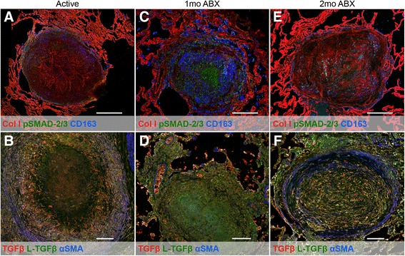
Fig. 3.

Tuberculous granulomas bear signs of TGFβ-driven fibrosis. Top panels feature granulomas stained for collagen I (red), phosphorylated SMAD-2/3 (green), and CD163 (blue). Bottom panels feature granulomas stained for TGFβ (red), L-TGFβ (green), and αSMA (blue). Magnification ×200. Scale bar represents 500 μm. a, b Granulomas from animals with active disease. c, d Granulomas from animals after 1 month of TB chemotherapy. e, f Granulomas from animals after 2 months of TB chemotherapy
