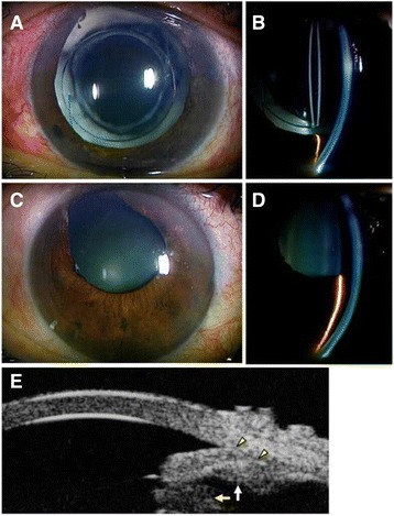
Fig. 1.

Slit lamp photographs. In the left eye, the pupil was markedly dilated, and a patent peripheral iridectomy and Soemmering’s ring were found (a, b). In the right eye, the anterior chamber was shallow despite a patent iridectomy. In the left eye, the anterior chamber was deep in the central region, but very shallow in the periphery, with a plateau-like iris configuration (c, d). Ultrasound biomicroscopy (UBM) showed a plateau-like iris and co-localized dilated pupil margin (between yellow arrow heads) with an enlarged Soemmering’s ring (yellow arrow) and anterior insertion of ciliary process (white arrow) (e)
