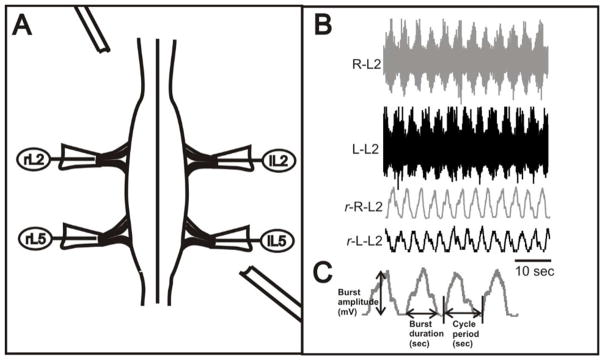
Figure 1. Experimental setup.
A: Suction recording electrodes were placed to monitor motor activity from ventral root nerve before, during and after perfussion of drugs. B: Extracellular recordings from rL2, and lL2 ventral roots after application of 6μM NMDA and 9μM 5-HT and 18μM dopamine (DA), showing locomotor-like activity characterized by left–right alternation in a P2 spinal cord. Raw (upper two traces) and rectified (lower two traces) recordings are shown. C: Representative segment of a rectified and smoothed trace from an actual control ventral root recording showing the locomotor-related parameters and how there were measured.

