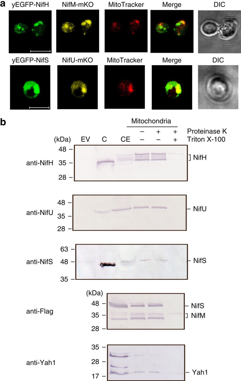
Figure 1. Mitochondrial targeting of Nif proteins.
(a) Confocal microscopy images of S. cerevisiae strains carrying synthetic sod2-yEGFP-nifH and sod2-nifM-mKO (strain GF5) or sod2-yEGFP-nifS and sod2-mKO-nifU genes (strain GF7), which expression was induced by galactose in the growth medium. The mitochondria of galactose-induced cells were localized with MitoTracker Deep Red. Scale bar, 5 μm. (b) Immunoblot analysis of isolated mitochondria developed with antibodies against NifH, NifU, NifS, Yah1 or FLAG (to detect NifS and NifM at the same time). EV represents cell-free extracts from recombinant yeast carrying pESC-His and pESC-Ura plasmids. CE represents cell-free extracts from recombinant yeast carrying NifH, NifM, NifU and NifS cloned into pESC-His and pESC-Ura plasmids (strain GF8). C represents control lanes with purified Nif proteins from A. vinelandii. Isolated mitochondria were treated with proteinase K either in the absence of detergent (for the removal of outer membrane proteins) or in the presence of Triton X-100 (to permeabilize the organelle).

