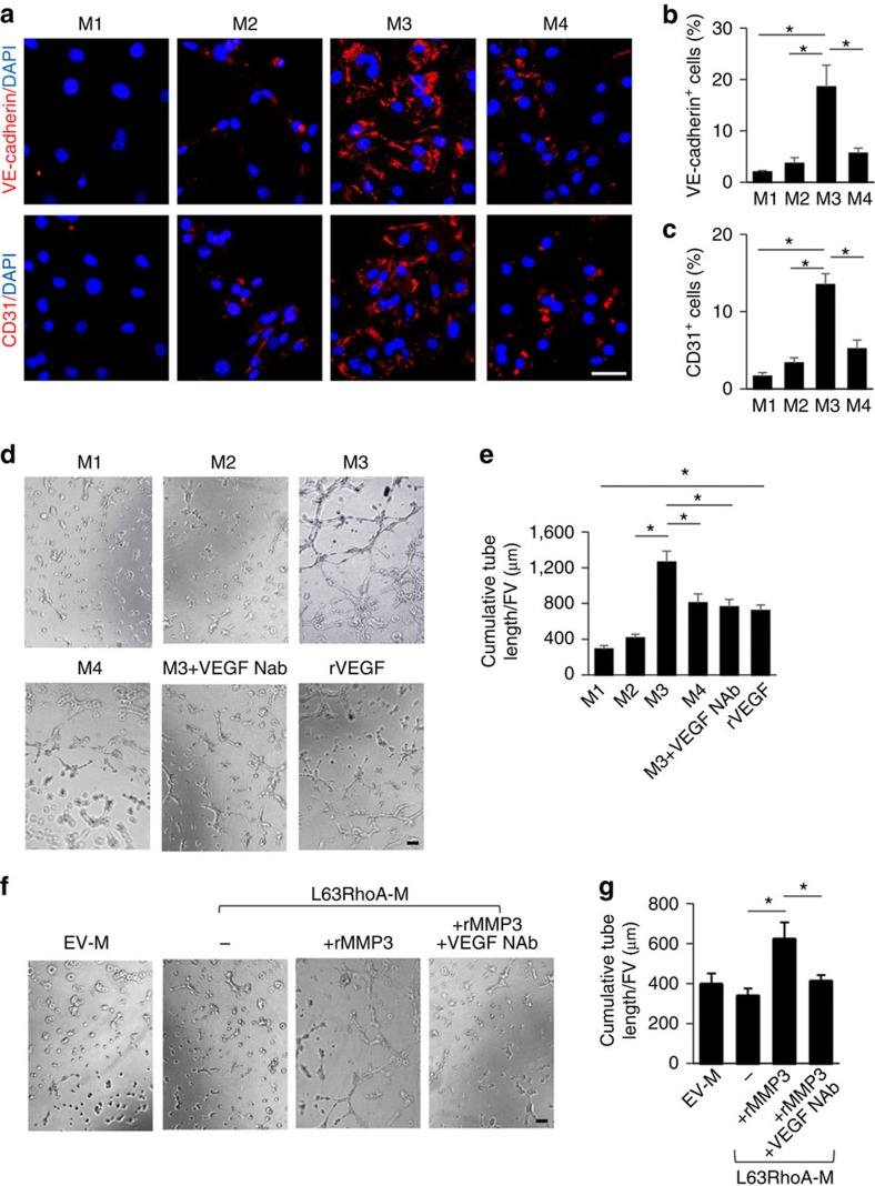
Figure 7. Inactivation of RhoA promotes endothelial differentiation of MSCs through MMP3.
Mouse MSCs were incubated with different media: αMEM with 1% serum only (M1), adding TGFβ1 (M2), adding both TGFβ1 and RhoA/ROCK inhibitor Y27632 (M3) or adding TGFβ1, Y27632 and a specific MMP3 inhibitor UK370106 (M4) for 6 days. Immunofluorescence staining of the cells using individual antibodies against VE-cadherin and CD31 (a). Quantification of the percentages of VE-cadherin- (b) and CD31- (c) positive staining cells out of total cells. n=5. Scale bars, 50 μm. Representative images of tube formation (d) and quantitative data of cumulative tube length (e) of MSCs cultured with indicated overlay medium (see the description in a). rVEGF treatment serves as a positive control. n=5. Representative images of tube formation (f) and quantitative data of cumulative tube length (g) of MSCs cultured with indicated overlay medium. n=5. Scale bars, 200 μm. Data are represented as mean±s.e.m. *P<0.01 as determined by ANOVA (b,e,g). DAPI, 4,6-diamidino-2-phenylindole.

