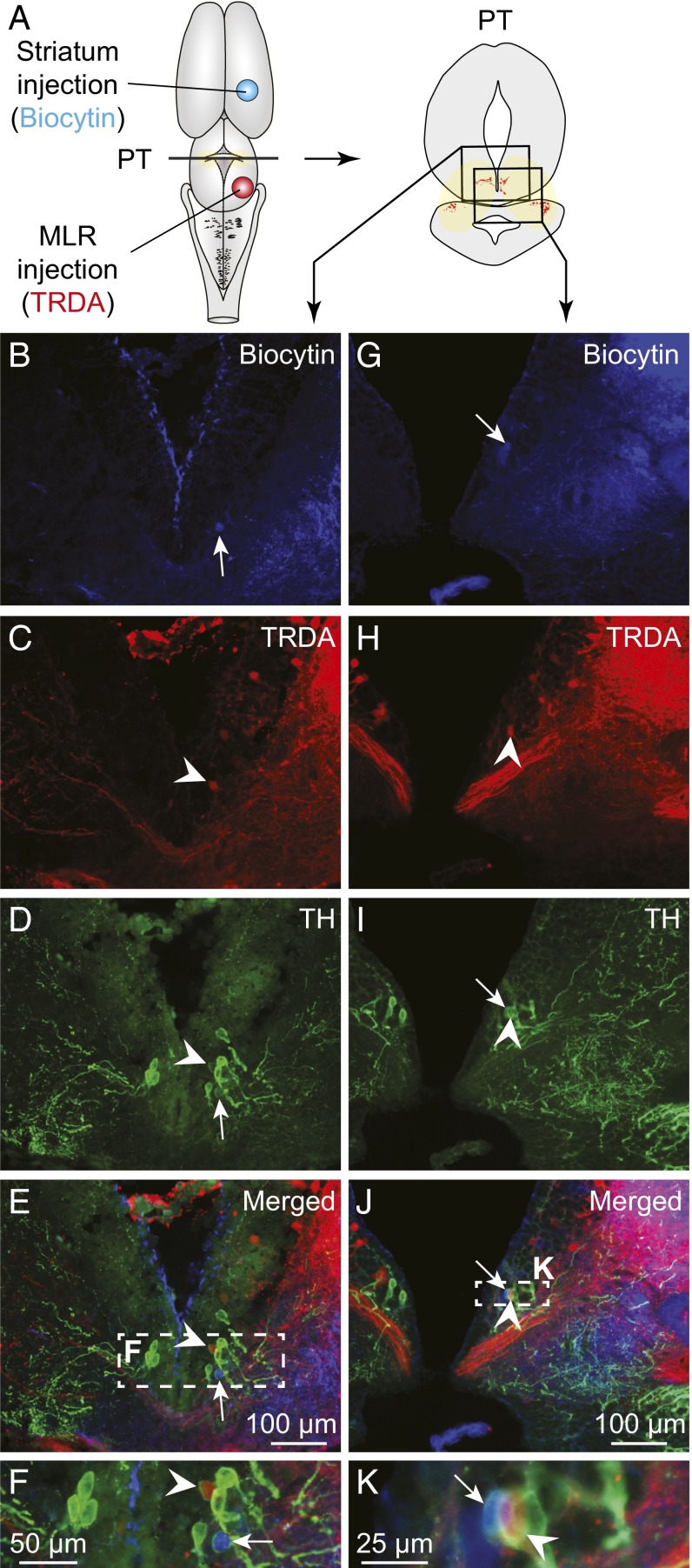
Fig. 2.
TH+ cells projecting to the MLR or to the striatum are intermingled in the PT. (A) Schematic dorsal view of the salamander brain, with the approximate location of the tracer injection sites in the striatum (biocytin, blue) and in the MLR (TRDA, red) (injection sites in Fig. S2 E and I, respectively). (Right) A diagram illustrates a PT cross section with the approximate location of the photomicrographs shown in B–E and G–J, obtained following triple-labeling experiments performed in two different animals. (B and G) Unilateral injection of a tracer in the striatum- (biocytin, blue) labeled cells in the PT. (C and H) Unilateral injection of another tracer in the MLR (TRDA, red) also labeled cells in the PT. (D and I) DA neurons of the PT were identified using immunofluorescence against TH (green). (E, J, F, and K) The photomicrographs were merged to show the three markers. Arrowheads indicate examples of TH+ cells of the PT that project to the MLR. White arrows point to TH+ cells projecting to the striatum. (F and K) Magnifications of the dashed rectangles in E and J, respectively.

